Spinal synovial cyst is a benign cystic mass originated from the facet joints. It is usually associated with degenerative spondylosis or spondylolisthesis and occurs frequently in lumbar region [1,2]. It can be asymptomatic or cause radiculopathy and symptoms associated with spinal stenosis. Magnetic Resonance Imaging (MRI) is the best diagnostic tool which demonstrates the nature and localization of the cyst and neural compression if it is present. However very rarely synovial cysts contain gas best seen on Computed Tomography (CT) [3,4,5,6]. In this report, we present a case of lumbar synovial cyst containing gas. Differential diagnosis of degenerative spinal cyst and intraspinal gas are also discussed.
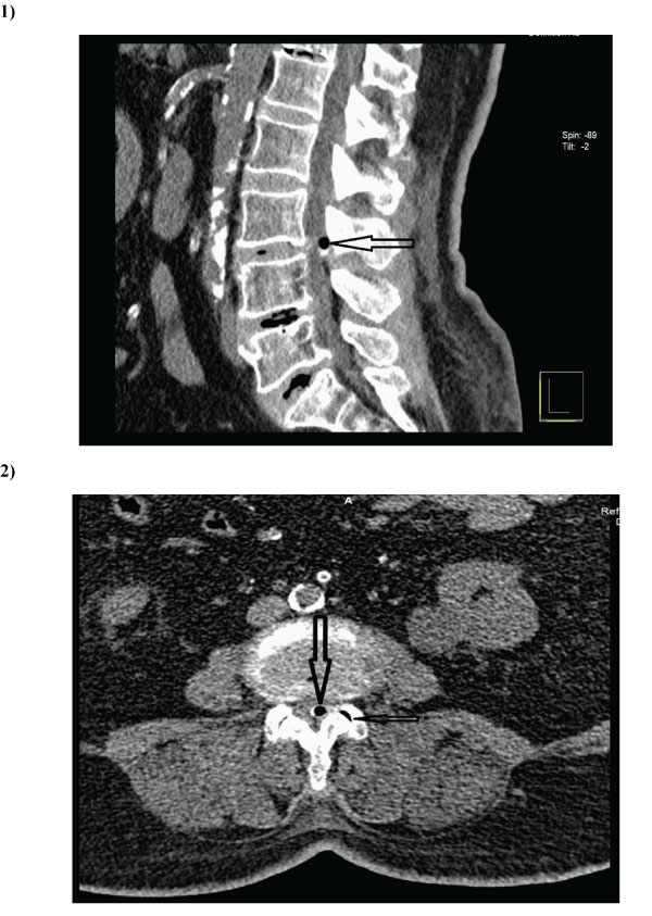
68-year-old women was admitted to the hospital with a low back pain radiating to her left leg for a week. She also complained of difficulties on walking for two years. On neurological examination, straight leg raising was positive at 45o on left. There was also a weakness of left knee extension with L4 hypoesthesia. Lumbar MRI showed a round hypointense lesion on both T1 and T2 weighted image adjacent to the left facet joint. There was no contrast enhancement following intravenous administration of contrast material. MRI appearance of the lesion suggested a calcified mass. On CT, a gas filled round synovial cyst with partially calcified wall was seen (Fig.1,2). Adjacent facet joint has also a vacuum sign. The patient underwent microsurgical removal of synovial cyst via L3 hemilaminotomy. A round mass with calcified wall compressing left L3 root removed. The wall of mass was not compressible due to calcification. All peripheral ligamentum flavum was also removed. Postoperative period was uneventful and symptoms were fully resolved in two months. Histopathological examination resulted in the diagnosis of synovial cyst.
Spinal degenerative cyst can arise from discs, facet joints, ligamentum flavum and posterior longitudinal ligaments [1]. Cyst of intervertebral discs and posterior longitudinal ligament are very rare and located in the ventral, ventrolateral spaces. Their diagnosis can be established intraoperatively. Classically, facet joints cysts include synovial cysts and ganglion cysts [2]. Synovial cysts develop from herniated synovial tissue. They contain xanthochromic joint fluid. Histologically they are lined by synovial epithelium and also have a connection with facet joint. Ganglion cysts occur secondary to mucoid degeneration of mesenchymal connective tissue. They don’t have synovial epithelium and contain mucoid gelatinous fluid. Histological appearance of ligamentum flavum cysts is similar to that of ganglion cyst. Today, ligamentum flavum and posterior longitudinal ligament cysts are also classified as facet cysts [1,2]. Spontaneous hemorrhages occur relatively more in ligamentum flavum cysts than others.
MRI is the best imaging technique for the diagnosis of degenerative spinal cyst. They usually appear as isointense lesions on T1 weighted images with a contrast enhanced rim while T2 weighted image show hyperintense mass [1,2]. Protein rich content due to hemorrhage can cause hyperintense appearance on both T1 and T2 weighted images. Differential diagnosis on MRI is usually on the basis of the location of degenerative cyst and may be difficult [1]. If there is a connection between the cyst and facet joint the diagnosis of synovial cyst is made while cysts distant from the joint are likely ganglion cysts.
Intraspinal gas or air collection is rare and is usually associated with vacuum phenomenon [3,4]. Vacuum phenomenon is the term defines the presence gas or air in joint spaces such as intervertebral discs, facet joints. Distraction of joints creates expansion of joint space and negative pressure. Dissolved nitrogen in tissues sublimates into gaseous state and occupies the joint space.
However, degenerative spinal cysts containing gas are very rare [3,4,5,6]. In our case left L3-4 facets also had vacuum phenomenon, so there was a connection between the cyst and the facet, confirming that the cyst is of synovial origin. MR is superior to CT in the diagnosis of synovial cyst. However, synovial fully gas filled cyst is better scan on CT because the gas appears as a signal void lesion on MRI, suggesting calcification. In the most of the gas containing synovial cyst, gas exists as a bubble within the fluid therefore, MRI usually shows the fluid nature of the lesion. On MRI the fully gas filled cysts appear as a signal void lesions suggesting calcification as in our case .Contrast enhancement of the cyst wall is a common radiological finding although calcified cyst walls do not show contrast enhancement .In our case ,as well as some other reported cases of MRI era ,the presence of gas within the synovial cyst was confirmed only after the CT examination [3,4,5,6].
Management of lumbar synovial cyst depends on patient’s clinical condition. Asymptomatic cases should be treated conservatively because spontaneous regression may occur [7]. In the patients with radicular symptoms and neurologic deficits, synovial cysts should be excised. Cyst removal via unilateral approach is usually sufficient. If the cyst is associated with spondylolisthesis or lumbar canal stenosis posterior, decompression and stabilization are recommended. Gas containing cysts could not cause different symptoms however, some authors thought that gas-containing cysts hardly regress and surgery is superior in the treatment of these cysts [3]. Percutaneous CT Guided aspiration of cyst contents is another treatment option although recurrence of gas containing cysts has been reported [8].

Figure 1 and 2: Sagittal (1) and axial (2) CT scan slices show an air filled intraspinal mass with partially calcified wall at L3-4 level (big arrows). On axial slice, vacuum sign in the adjacent facet joint is seen (small arrow).
Synovial cysts arise from herniated synovial tissues of facet joints of spine. They usually occur in lumbar region and contain xanthochromic fluid. However, synovial cysts rarely contain air or gas. Although, MRI is the best imaging technique in the diagnosis of spinal degenerative cysts, intraspinal air is better seen on CT. Presence of air within the degenerative cyst confirms the synovial origin of the cyst. Symptomatic gas containing synovial cysts should be excised, because these cysts very rarely regress spontaneously.